
Radiography & Ultrasonography
Here at Grange Veterinary Surgery, we use both methods frequently depending on our clinical suspicions, as one method may be more suitable to an investigation than the other. Occasionally we need to use a number of investigative methods.
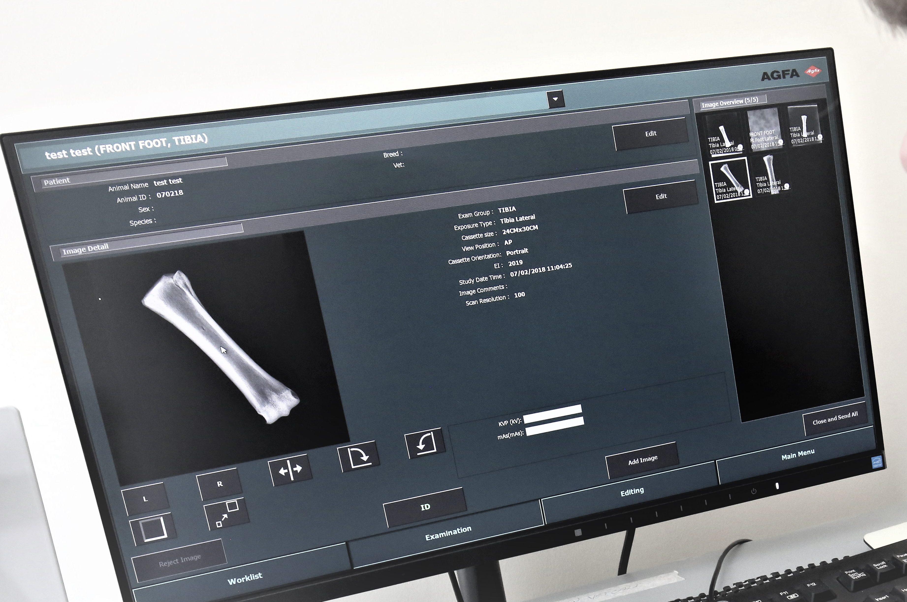
Radiography (X-ray)Radiography is commonly used to assess lameness and painful joints, heart and lung disorders, vomiting and digestive problems and urinary issues. It is performed with either a general anaesthetic or a sedative and usually within a day to allow you to take your pet home once the procedure is over.
We have a modern digital x-ray suite which is second to none.
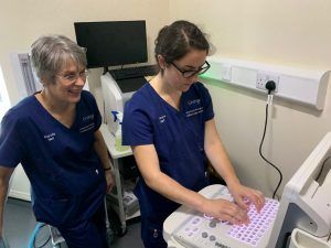
Ultrasonography (ultrasound)Ultrasonography is another painless and non-invasive method of investigation. Ultrasonography is commonly used for investigating liver, spleen, bladder or reproductive disorders. Its use in diagnosing gastrointestinal and cardiac issues is increasing. We also use ultrasonography to guide tissue biopsies. Frequently ultrasound images can be generated from a conscious patient. Occasionally a sedative is required.